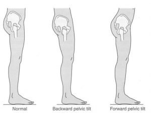
FUNCTIONAL ANATOMY
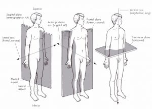
Anatomical Terms and Planes
Anterior: towards or at the front of the body.
Posterior: towards or at the rear of the body.
Superior: toward the head / upper part of a structure.
Inferior: away from the head or towards the lower part of a structure.
Lateral: away from the midline of the body, on the outer side.
Medial: towards the midline of the body, on the inner side.
Proximal: closer to the trunk of the body part.
Distal: further from the trunk of a body part.
Superficial: towards or at the surface.
Deep: away from the body surface.
Sagittal plane: runs anteriorly-posteriorly, divides body into left and right.
Frontal (coronal) plane: runs laterally, divides body into front and back.
Transverse plane: runs horizontally across body, divides into upper and lower.
Movement Definitions
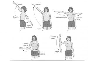
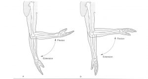
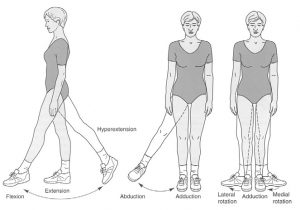
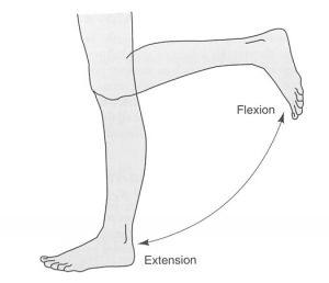
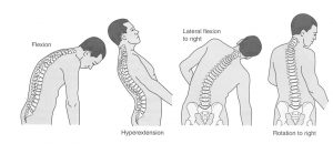
Flexion: movement that decreases joint angle.
Extension: movement that increases joint angle.
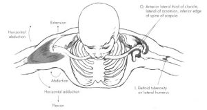
Abduction: movement of limb away from midline of body.
Adduction: movement of limb towards midline.
Circumduction: combination of all of the above performed in succession (draws cone shape).
Horizontal Abduction / Extension: movement away from the midline / increasing the joint angle in the horizontal plane.
Horizontal Adduction / Flexion: movement towards the midline / decreasing the joint angle in the horizontal plane.
Medial Rotation: movement around long axis of bone towards midline.
Lateral Rotation: movement around long axis of bone away from midline.
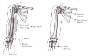
Supination: movement of forearm so that palm faces anteriorly (turning palm up).
Pronation: movement of forearm so that palm faces posteriorly (turning palm down).
Inversion: sole of foot turned medially.
Eversion: sole of foot turned laterally.
Plantarflexion: toes move away from lower leg / extension of ankle (point toes).
Dorsiflexion: toes move towards lower leg / flexion of ankle (pull toes up).
Protraction: movement anteriorly in transverse plane.
Retraction: movement posteriorly in transverse plane.
Elevation: moving superiorly in frontal plane (lifting).
Depression: moving inferiorly in frontal plane (dropping).
Muscle actions
agonist – A muscle or muscle group that is described as being primarily responsible for a specific joint movement when contracting.
antagonist – A muscle or muscle group that counteracts or opposes the contraction of another muscle or muscle group.
synergist – Muscles that assist in the action of the agonists but are not primarily responsible for the action; known as guiding muscles, they assist in refined movement and rule out undesired motions.
stabilizers – Muscles that surround the joint or body part and contract to fixate or stabilize the area to enable another limb or body segment to exert force and move; known as fixators, they are essential in establishing a relatively firm base for the more distal joints to work from when carrying out movements.
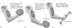
Types of contractions
Understanding of the different ways a muscle can contract is important when analyzing movement and exercise. The 3 forms of muscle contraction are listed below:
Concentric contraction
The muscle develops tension while it shortens. The muscle develops enough force to overcome the resistance. This occurs during the main exertive phase of an exercise such as the push up in the bench press or the standing up from a squat.
Eccentric contraction
The muscle lengthens under tension. This allows the muscle to control the lengthening of a movement and prevent injury. Eccentric contractions control movements occurring with gravity or resistance. An example of an eccentric contraction would be lowering the weight during the bench press.
Isometric contraction
The muscle develops tension but the muscle length doesn’t change. This would occur if you hold a squat position against a wall.
A good trainer will continue to learn, educate themselves and be a role model to others.
Functional Anatomy of The Major Joints
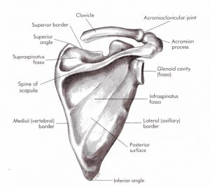
Scapula
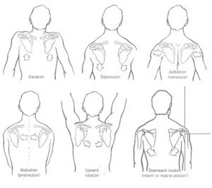
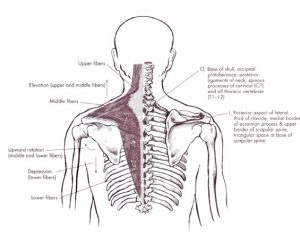
Trapezius
Action
Upper: elevation of scapula, extension of the head.
Middle: elevation, lateral rotation and retraction of scapula.
Lower: depression, retraction and lateral rotation of scapula
Levator Scapulae
Action
Elevates scapula.
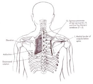
Rhomboid Major
Action
Retraction of scapula.
Medial rotation of scapula.
Elevation of scapula (during retraction).
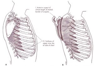
Serratus Anterior
Action
Protraction of scapula.
Lateral rotation of scapula (lower fibers, slight).
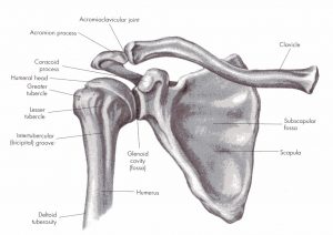
Glenohumeral Joint
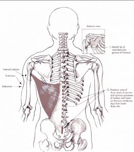
Latissimus Dorsi
Action
Adduction of glenohumeral joint Extension of glenohumeral joint Internal rotation of glenohumeral joint.
Horizontal abduction of glenohumeral joint.
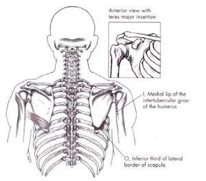
Teres Major
Action
Adduction of glenohumeral joint.
Extension of glenohumeral joint.
Internal rotation of glenohumeral joint.
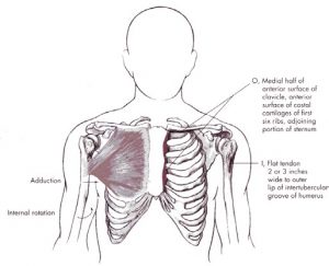
Pectoralis Major
Action
Upper: horizontal adduction, flexion, internal rotation of glenohumeral joint.
>90o abduction: assists in abduction.
<90o abduction, assists in adduction.
Lower: horizontal adduction internal rotation, extension and adduction of glenohumeral joint.
Deltoid
Action
Anterior: flexion of glenohumeral joint, internal rotation.
Medial: abduction of shoulder joint.
Posterior: extension, horizontal abduction, external rotation of glenohumeral joint.
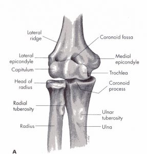
Elbow Joint
(A- Anterior, B- Lateral and C- Medial Views)
Biceps Brachii
Action
Flexion and supination of elbow joint.
Flexion of shoulder joint.
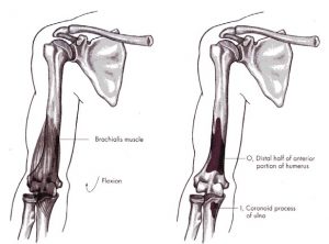
Brachialis
Action
Flexion of elbow joint.
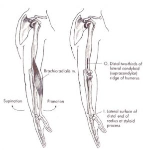
Brachioradialis
Action
Flexion of elbow joint.
Pronation from supinated to neutral.
Supination from pronated to neutral.
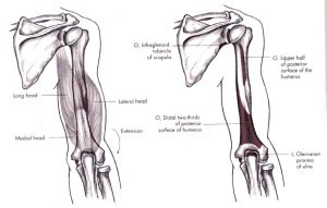
Triceps Brachii
Action
Extension of elbow joint.
Long head; extension and adduction of shoulder joint.
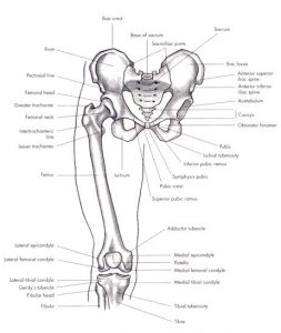
Pelvis and Femur (Anterior View)
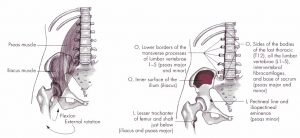
Iliopsoas
Action
Flexion of hip.
External rotation of femur.
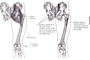
Gluteus Maximus
Action
Extension and external rotation of hip.
Inferior fibers adduct hip.
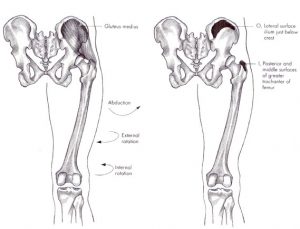
Gluteus Medius
Action
Abduction of hip.
Posterior fibers: external rotation as abduction occurs.
Anterior fibers: internal rotation.
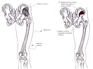
Gluteus Minimus
Action
Abduction of hip.
Internal rotation as abduction occurs.
Biceps Femoris
Action
Extension of hip Flexion of knee.
External rotation of hip.
External rotation of flexed knee.
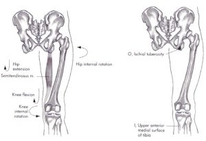
Semitendinosus
Action
Extension of hip Flexion of knee.
Internal rotation of hip.
Internal rotation of flexed knee.
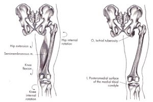
Semimembranosus
Action
Extension of hip.
Flexion of knee.
Internal rotation of hip.
Internal rotation of flexed knee.
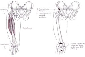
Rectus Femoris
Action
Flexion of hip.
Extension of knee.
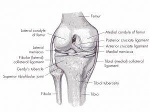
Knee Joint
(Anterior, Posterior and Superior Views)
Rectus Femoris
Action
Flexion of hip.
Extension of knee.
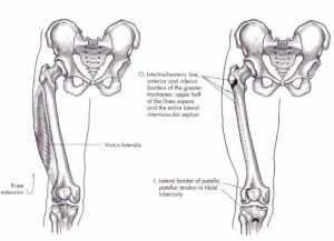
Vastus Lateralis
Action
Extension of knee.
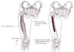
Vastus Intermedius
Action
Extension of knee.
Vastus Medialis
Action
Extension of knee.
Tibia, Fibula and Foot
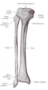
Movements at the ankle joint
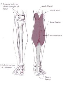
Gastrocnemius
Action
Plantar flexion of ankle.
Flexion of knee.
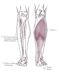
Soleus
Action
Plantar flexion of ankle.
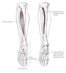
Tibialis Anterior
Action
Dorsiflexion of ankle.
Inversion of foot.
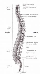
Spine
Erector Spinae
Action
Extension of spine (bi-lateral).
Lateral flexion of spine (uni-lateral).
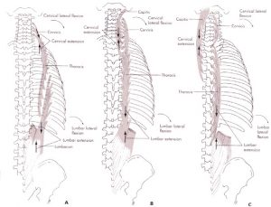
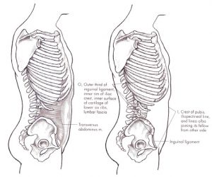
Transverse Abdominus
Action
Abdominal compression.
Stabilization of lumbar spine.
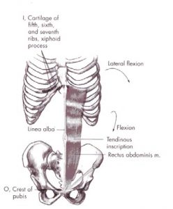
Rectus Abdominus
Action
Flexion of spine.
Lateral flexion (uni-lateral).
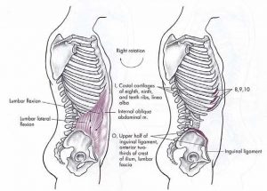
Internal Oblique’s
Action
Right & Left Rotation.
Right & Left Lateral Flexion.
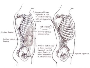
External Obliques
Action
Right & Left Rotation.
Right & Left Lateral Flexion.
Special Comment on the transverse Abdominus
The transverse abdominus muscle (also known as the transversus abdominus or trans abs for short) has been in the headlines over the last few years because of its role as a core stability abdominal wall muscle in the treatment for lower back pain.
The transverse muscle is a corset-like abdominal wall muscle that is the deepest (or the furthest in) of the abdominal muscles. It is attached to the body in a few places including the spine, the ribs and the pelvis. It runs in a circular, tube like fashion around the whole of the trunk.
It is one of several abdominal muscles that have a role to play in treatment for lower back pain. These others include the internal and external oblique’s and also the rectus abdominis.
Many people don’t realize that some abdominal wall muscles actually attach at the spine and pelvis and create a tube like support structure for your back. This is why they play an important role in giving your back support and why they have come into such prominence in the treatment for lower back pain.
Commonly people tend to think of abdominal muscles as being the six pack muscles you get when you do a lot of sit ups. Actually, sit ups mostly focus on the rectus abdominis muscles and also to a lesser degree the internal and external oblique’s.
Basic Transverse Exercise – TRY IT! Setup
Try standing first. Once you’ve mastered that, do the exercise on all fours for greater resistance.
Execution
- Exhale deeply.
- Towards the end of your exhale, clench your abdominal muscles such that your belly button moves towards your spine (i.e. backwards) a half inch or so. Do not suck in your gut; that’s a diaphragm exercise.