STRUCTURE & FUNCTION
The Musculo-skeletal system
Introduction
A basic level of knowledge of the structure and function of the musculo-skeletal system is essential to have a better understand of body movement control and thereby more effectively on how to improve strength, endurance and flexibility.
This section should enhance your knowledge of the structure and function of the major muscles, bones and joints of the human body.
In other sections you will analyse how your body coordinates movement, and then use this information to ensure your exercise participants perform activities that are appropriate, safe, efficient and enjoyable.
Quick Points:
- The skeleton comprises 206 bones, although human variation may account for several absent or additional bones.
- Approximately 650+ muscles comprise close to 40% of the overall body mass.
- The human body comprises approximately 10,000,000,000 nerve fibers that are distributed over 100,000 km.
The major systems coordinating body movement
The skeleton comprises 206 bones, although human variation may account for several absent or additional bones.
The skeletal system
Comprises bones and associated soft tissues that form the joints of the body. It is the supporting framework, but with other functions too, for example, blood production, calcium reservoir.
The muscular system
Comprises various muscle fiber types, shapes and arrangements. The attachments to bones allow contractions to produce movements by pivoting at the joints.
Muscles respond to signals from the brain, which travel along the nerves and cause each muscle fiber to contract.
The nervous system
Comprises brain, spinal cord and peripheral nerves. This system controls and coordinates the muscles as well as many other body activities. The sensory component of the nervous system uses the five senses: hearing, sight, taste, smell and touch to analyse the nature of conditions around the body. The motor part then controls the muscles and enables movement to occur.
In summary
The body is therefore a system of levers = BONES. The junctions between these are the JOINTS that are the points around which the MUSCLES provide the force to produce MOVEMENT under the control of the nervous system.
The Skeletal System
Major functions
SUPPORT — vertebral column, pelvis
PROTECTION — skull, rib cage, pelvis
MOVEMENT — spine, limbs
Functions of the Skeleton
The skeleton performs several important functions.
Support
Acts as a framework for the body giving support to soft tissues and providing points of attachment for muscles.
Movement
Through providing anchor points for muscles, allows movement of body parts.
Protection
Protects many of the vital organs:
- Skull- brain
- Vertebrae- spinal cord
- Ribs- heart and lungs
- Pelvis- digestive system and reproductive organs (female)
Storage of Minerals
Calcium, sodium and potassium are stored in bones.
Blood Cell Production (Haemopoesis)
Red blood cells are produced in the marrow of the long bones.
Components
- Bones
- Joints
Bones
The skeleton comprises:
- Groups of long bones e. those making up the arms and legs, involved primarily in movement.
- The vertebrae of the spine and the bones of the pelvis for central support.
- The rib cage and skull that protect vital organs.
- The short or cuboidal bones of the wrist and ankle.
Structure of Bone
Bone is composed of a number of different components.
Periosteum
- Fibrous sheath that covers bones.
- Contains the blood vessels and nerves that provide nourishment and sensation to the bone.
- Also contains osteoblasts that assist in repair of the bone after injury.
Compact Bone
- Hard dense bone located in the shaft of long bones and around the end of spongy bone at joints.
- Short bones composed entirely of this.
Spongy Bone
- Porous bone located at the end of long bones, in vertebrae and flat bones
- Allows strength with a reduction in weight.
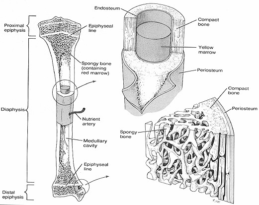
Epiphysis
- Bulbous end of long bone containing spongy bone.
- Covered by articular cartilage to form joints.
Diaphysis
- Long middle portion of bone connecting epiphyses.
Medullary Cavity
- Hollow cavity in diaphysis containing bone marrow.
Marrow
- Red bone marrow produces red blood cells.
- Yellow bone marrow acts as storage for fat.
Bone Growth
Because bone is made up of minerals and is hard, many people think that it is not living material. But a bone in a living animal consists of both living tissue and non-living substances. Within the ‘alive bone’ there are blood vessels, nerves, collagen, and living cells including: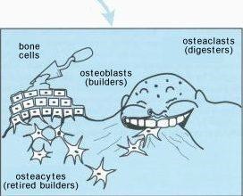
- osteoblasts (cells that help form bone), and
- osteoclasts (cells that help eat away old bone).
Bone also contains cells called osteocytes, which are mature osteoblasts that have ended their bone-forming role. These cells engage in metabolic exchange with the blood that flows through the bones. The nonliving, but very important, substances in bone are the minerals and salts.
In the fetus, most of the skeleton is made up of cartilage, a tough, flexible connective tissue that has no minerals or salts. As the fetus grows, osteoblasts and osteoclasts slowly replace cartilage cells and ossification begins.
Ossification is the formation of bone by the activity of osteoblasts and osteoclasts and the addition of minerals and salts. Calcium compounds must be present for ossification to take place. Osteoblasts do not make these minerals, but must take them from the blood and deposit them in the bone. By the time we are born, many of the bones have been at least partly ossified.
In long bones, the growth and elongation (lengthening) continue from birth through adolescence. Elongation is achieved by the activity of two cartilage plates, called epiphyseal plates, located between the shaft (the diaphysis) and the heads (epiphyses) of the bones (Figure 5). These plates expand, forming new cells, and increasing the length of the shaft. In this manner, the length of the shaft increases at both ends, and each head of the bone moves progressively apart. As growth proceeds, the thickness of the epiphyseal plates gradually decreases and this bone lengthening process ends. In humans, different bones stop lengthening at different ages, but ossification is fully complete by about age 25. During this lengthening period, the stresses of physical activity result in the strengthening of bone tissue.
In contrast to the lengthening of bone, the thickness and strength of bone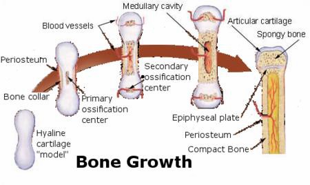 must continually be maintained by the body. That is, old bone must be replaced by new bone all the time. This is accomplished as bone is continually deposited by osteoblasts, while at the same time, it is continually being reabsorbed (broken down and digested by the body) by osteoclasts. Osteoblasts are found on the outer surfaces of the bones and in the bone cavities. A small amount of osteoblastic activity occurs continually in all living bones (on about 4% of all surfaces at any given time) so that at least some new bone is being formed constantly. Normally, in fact, except in growing bones, the rates of bone deposition and absorption are equal to each other so that the total mass of bone remains constant.
Usually, osteoclasts exist in small but concentrated masses, and once a mass of osteoclasts begins to develop, it usually eats away at the bone for about three weeks, eating out a tunnel that may be as large as 1 millimeter in diameter and several millimeters in length. At the end of this time the osteoclasts disappear and the tunnel is invaded by osteoblasts instead; then new bone begins to develop, Bone deposition then continues for several months, the new bone being laid down in successive layers of concentric circles on the inner surfaces of the cavity until the tunnel is filled. Deposition of new bone ceases when the bone reaches the surface of the blood vessels supplying the area. The canal through which these blood vessels run, called the haversion canal, therefore, is all that remains of the original cavity. This process continues until about age 40, when the activity of osteoblasts slows and bones become more brittle. Let’s look at some important factors that are necessary to produce healthy bone.
Bone development is influenced by a number of factors, including nutrition, exposure to sunlight, hormonal secretions, and physical exercise. For example, exposure of skin to the ultraviolet portion of sunlight is favorable to bone development, because the skin can produce vitamin D when it is exposed to such radiation. Vitamin D is necessary for the proper absorption of calcium in the small intestine. In the absence of this vitamin, calcium is poorly absorbed, the bone matrix is deficient in calcium, and the bones are likely to be deformed or very weak. Vitamins A and C also are needed for normal bone growth and development.
There are many causes of osteoporosis:
- the lack of physical stress on the bones because of less activity;
- malnutrition, including not enough calcium in the diet, to the extent that sufficient bone matrix cannot be formed;
- lack of vitamin C, which is necessary for osteoblasts to create healthy bone matrix;
- the lack of estrogen secretion, which occurs in women after menopause (estrogen is also important to stimulate osteoblast activity); end,
- old age, when the body simply cannot form sufficient bone matrix.
Classification of Bones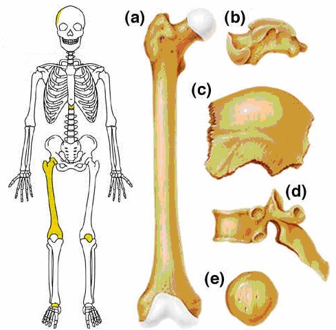
Bones can be classified according to their shape.
Long Bones (a)
- Longer than they are wider.
- Have long axis.
- Humerus, femur, radius, ulna.
Short Bones (b)
- Roughly cube shaped.
- Do not have long axis.
- Carpals in wrist, tarsals in feet
Flat Bones (c)
- Thin bone with greater surface area for protection.
- Cranial bones, ribs, sternum.
Irregular Bones (d)
- Various shapes that don’t fit other categories.
- Vertebrae.
Sesamoid Bones (e)
- Shaped like a sesame seed.
- Short bones that form within a tendon eg patella.
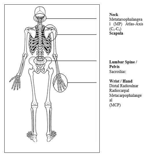
Major Bones of The Body

JOINTS
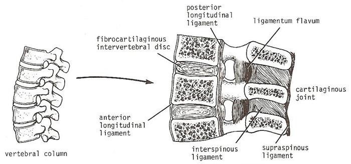
A joint is the point where two or more bones meet. There are three main types of joints; Fibrous (immoveable), Cartilagenous (partially moveable) and Synovial (freely moveable).
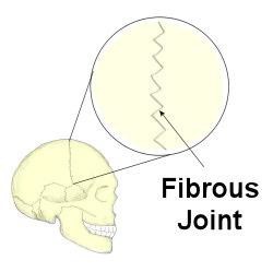
- Fibrous — fixed joints — don’t allow Mainly provide support and protection, for example, skull, pelvis, ribs. Above Image
- Cartilaginous — slightly movable joints-allow a small amount of movement & are mostly supporting in function, eg. between vertebra. See below Image
.
3. Synovial – movable joints – allow free movement
Synovial joints are the most common in the body. They are more structurally complex than cartilaginous or fibrous joints, and are basically fluid-filled cavities that allow minimal friction between the articulating surfaces. All synovial joints have a similar structure, but the shape of the articulating surfaces varies, and this determines the type and extent of motion at each joint.
The bone ends are covered with smooth articular cartilage and are connected by an enclosing joint capsule. The capsule is lined with a smooth synovial membrane that secretes synovial fluid to lubricate and provide nutrition to the joint. The joint is strengthened by ligaments attached to adjacent joint margins.
Structure of Synovial Joints
Characteristics of Synovial Joints
Five structural characteristics are common to all synovial joints with three others found only in specific joints:
Fibrous Capsule
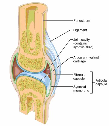
- Strong fibrous tissue envelope surrounding joint.
- Attaches to periosteum of bones.
- Main source of joint stability.
Synovial Membrane
- Layer covering inside of joint capsule.
- Does not covering articular surface of joint.
- Produces synovial fluid.
Articular Cartilage
- Smooth, white tough covering on surface of bone.
- Protects bone tissue and helps reduce friction during movement.
Synovial Fluid
- Yellowish, waxy fluid that fills the joint space in the joint capsule.
- Forms fluid cushion to assist in shock absorption.
- Lubricates articular surfaces.
- Provides nutrients for articular cartilage.
Ligament
- Strong fibrous bands that assist in maintaining joint stability.
- Attach bone to bone.
- Consist of non elastic collagen.
- Can be intra-articular (within joint capsule) or extra-articular (outside joint capsule).
Meniscus (often referred to as cartilage)
- Hard discs located inside joint capsule on articular surfaces.
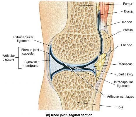
- Attached to joint capsule.
- Designed to absorb shock, help maintain stability, protect bony surfaces and direct flow of nutrients to high wear areas.
- Found in knee, wrist and jaw.
Fat Pad
- Small pads of fat located in crevasses around joint.
- Form protective cushioning for structures around joint.
- Found in knee joint.
Bursa
- Small fluid filled sacs located around joint in high friction areas.
- Particularly found where tendons lie in close proximity to bony surfaces.
- Found around hip joint, knee joint, elbow joint and shoulder joint.
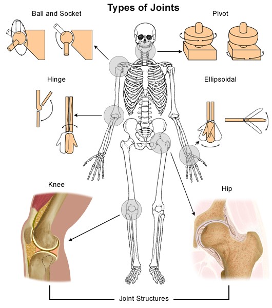
Common Types of Synovial Joints
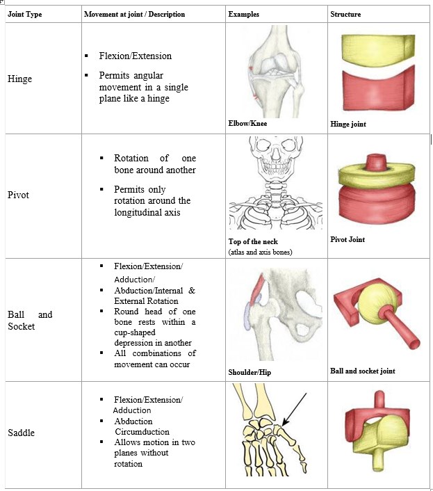
Factors affecting movement at a synovial joint
Bony configuration
The shape of the bones determines the direction a joint can move.
- Ball and socket joint — for example, hip or shoulder — a spherical head fits into a cup-like cavity and allows a large range of movement.
- Hinge joint — for example, elbow, knee — allows movement in one plane only, that is, in two directions, for example, flexion or extension.
- Pivot joint— for example, 1st and 2nd cervical vertebrae and radio — ulna joint of A bone rotates around a bony prominence on another bone.
- Gliding or sliding joint — for example, wrist, hands or feet — flat surfaces slide on each other.
The soft tissues
- Ligaments — are relatively inelastic bands of fibrous tissue which provide extra stability for the joint, for example, lateral ligament of ankle, ligaments of knee.
- Tendons — bands of inelastic fibers by which muscles insert into bone, also affect joint stability and movement.
- Muscles — the muscle action, plus its degree of extensibility, permits or restricts joint movement.
Major Joints of the Body

Spine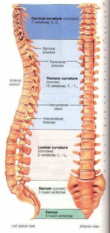
In today’s society we are often taught that the human spine is weak or vulnerable and that we should be very careful not to injure it. If look closely at the spine you can see how tough it is and how well designed it is to do its job. The spine is an amazingly strong and clever structure. However we do need to strive to protect the components of the spine, as over a period of time the wear and tear we place on it begins to affect its components. Let’s take a closer look at its components.
- Central axis of the body composed of 33 bones divided into 5 regions.
- Individual bones known as vertebra.
Components of Spine
Cervical, Thoracic and Lumbar Spine
Your spine is made up of 24 vertebrae. Each vertebra is made up of the vertebral body in the front, the facet joints in the back and the pedicles, which connect the vertebral body to the facet joints. The human spine is divided into three sections: 1) the cervical spine or neck is made up of 7 vertebrae, 2 – the altas and axis being C1 and C2 respectively) the thoracic spine made up of 12 vertebrae and 3) the lumbar spine or low back which consists of 5 vertebrae.
Curves of theSpine
Has 4 curves in sagittal plane named after area located
- Cervical curvature: curve is concave posteriorly.
- Thoracic curvature: curve is convex posteriorly.
- Lumbar curvature: curve is concave posteriorly.
- Sacral curvature: curve is convex posteriorly.
Curves designed to increase stability of structure and ability to absorb force.
Curves known as:
- Lordosis: lumbar and cervical.
- Kyphosis: thoracic.
- Scoliosis: lateral deviation (abnormal).
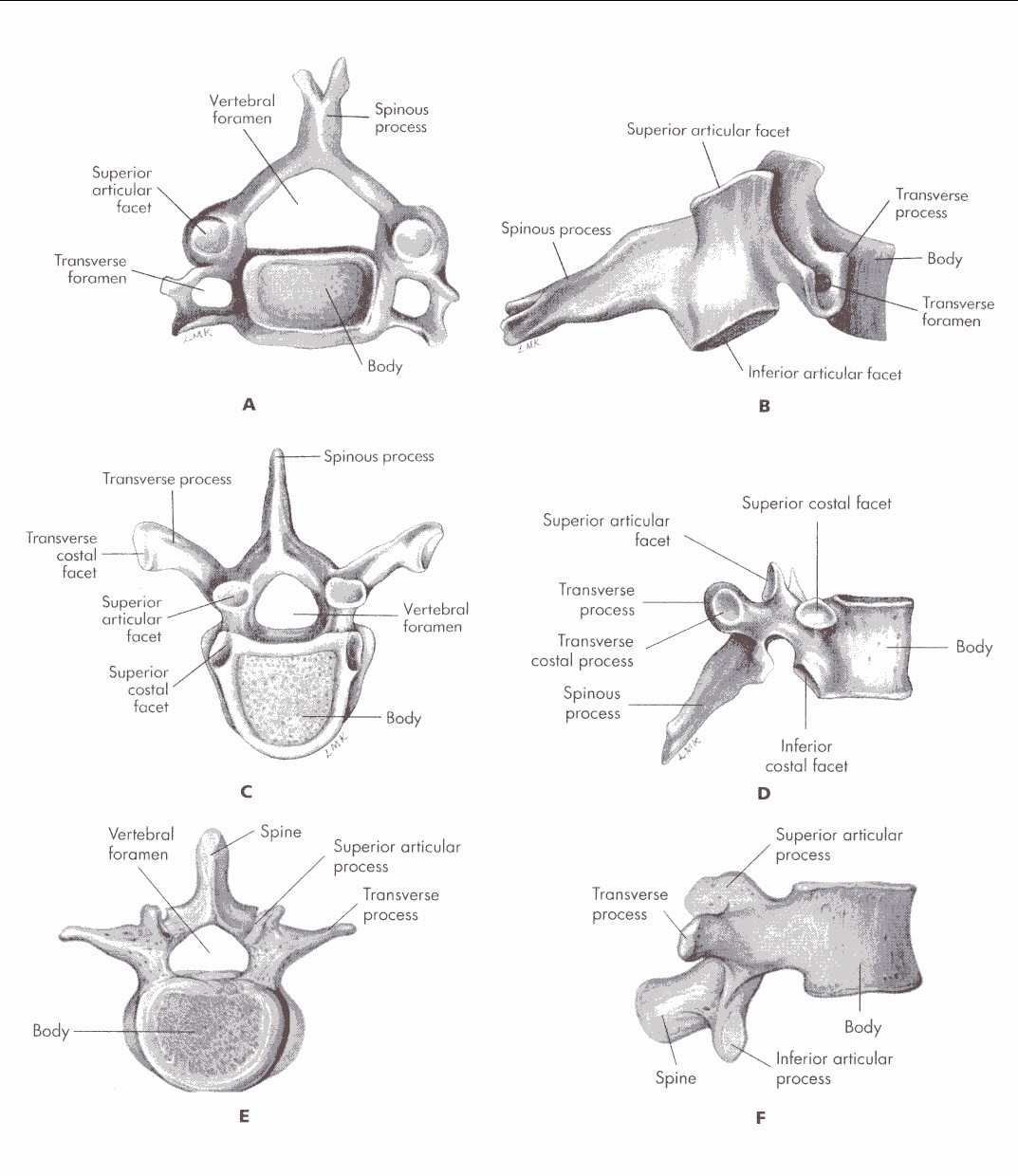
SpinalVertebrae
- Individual bony structures that make up the spine.
- Have a body (or central component) that transfers weight along the column.
Have bony projections called a process that allow attachment of ligaments and muscles.
- Spinous Process project posteriorly.
- Transverse Process project laterally.
- Have facet joints that allow movement of vertebra on each other.
- Protect the spinal cord via the spinal canal.
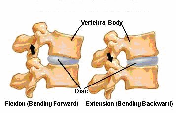
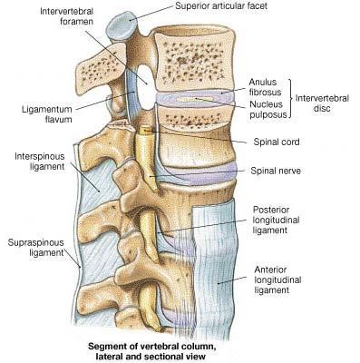
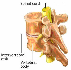
- Each vertebrae is separated by an intervertebral disc.
Cervical Vertebrae (A & B) Thoracic Vertebrae (C & D) Lumbar Vertebrae (E & F).
Intervertebral Joints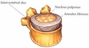
- The spine can be viewed as a series of bony blocks stacked on top of one another.
- In between each bony segment is a specialised structured called an intervertebral disc.
- The discs serve to act as a shock absorption system and allow movement of the spine.
- The tough outer layer of the disc is the Annulus Fibrosus.
- This annulus surrounds a soft, elastic and gelatinous core Nucleus Pulposus.
- There is minimal movement between each individual vertebrae otherwise the spinal cord would risk damage or damage to the nerves projecting from the side of the vertebrae.

- However as a functional unit , the spine is capable of a great range of movement.
- Numerous ligaments are attached to the bodies and processes of all vertebrae to bind them together and stabilize the vertebral column.
- While the joints between each vertebrae are classified as cartilaginous (slightly moveable), the vertebrae do articulate with one above and below via a plane type of synovial joint called a facet joint.